If you
disable the "Active Content" in your browser you may not
be able to view the animations or videos supplied in
this lab. If prompted you should "Allow Blocked
Content".
Access each of the listed
documents above and print them off. When you submit your
lab report you will need to compile all of the documents
listed above, stapled together in the order listed in
the table above. Sketches must be performed free hand
(not traced or copy and pasted). Sketches must be
performed using the printed links as given above. You
are not allowed to perform the sketches on blank sheets
of paper or lined sheets of paper. Sketches performed
without using these forms above will not be accepted.
You can use the MS WORD links to access the questions,
tables and charts in order to input your values or
answers electronically and then print them off when
finished to include with your lab report. Alternatively
you can print the questions, tables and charts forms out
and input your values or answers by hand. The PDF file
format will not allow you to input values or answers
electronically. Please collate and order the pages in
your lab report in the order they are listed in the
table above. The cover page is only available using the
PDF file format.
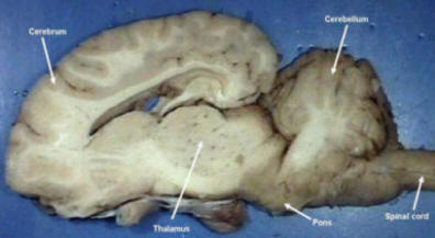
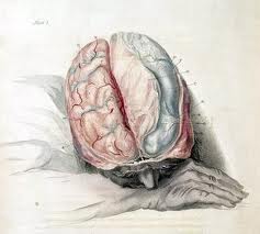
This section
of our study concerns the external and internal structure of the
brain. Comparisons will be made between the sheep brain and the
human brain. Once you have located a structure on the sheep
brain, try to find a comparable structure on the human brain.
Keep in mind that direct comparisons are not always possible.
SKETCH
1
**Using the
images presented in this lab book and your text book, sketch an
image of the sheep brain and identify and label the following:
Cerebrum, Cerebellum, Pons, Medulla oblongata, Midbrain
Use the links below to view a
tutorial on human and sheep brain anatomy.
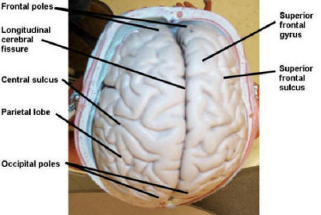
In both
the sheep brain and human brain, the cerebrum is the largest
portion of the brain. The cerebrum is divided along the median
by the longitudinal cerebral fissure line to form two cerebral
hemispheres. The surface of the cerebrum is covered with ridges
and furrows of varying depths. The deeper furrows are called
fissures and the shallow furrows are called sulci. The ridges or
convolutions are called gyri. A frontal section shows how this
in-folding of the cerebral surface increases the amount of gray
matter of the brain in a given space. Each half of the cerebrum
is divided into four lobes. The frontal lobe is the most
anterior portion. The occipital lobe is the most posterior lobe
of the brain. The temporal lobe lies on the lateral side of the
brain. An imaginary horizontal dotted line provides a border
between the temporal and parietal lobes.
Examine the surface of the cerebellum. Note
that its surface is furrowed with sulci. The human cerebellum is
constricted in the middle to form right and left
hemispheres. The cerebellum plays an important role in the
maintenance of posture and the
coordination of complex muscular movements.
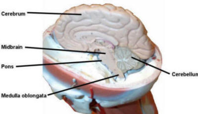
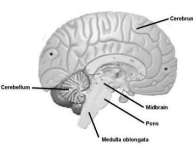
The remainder of the brain consists of
structures collectively referred to as the brain stem. This
lower portion of the brain includes the midbrain, diencephalon,
pons varolii, and medulla oblongata and excludes the cerebrum
and cerebellum.
Midbrain
Force the
cerebellum downward with the thumb to expose the midbrain. The
midbrain is closely associated with the sense of sight.
Portions of the midbrain are important analytical centers
concerned with brightness and sound discrimination.
Diencephalon
(Interbrain)
The pineal gland is a portion of the
diencephalon. There are indications that this gland is a remnant
of the third eye that exists in some reptiles. The hormone
melatonin is produced by the pineal gland. The precise role of
melatonin in humans is not completely understood. The
diencephalon is difficult to observe because it is in front of
the midbrain and lies below the cerebrum. The thalamus,
hypothalamus, and mammillary bodies are glands that are also
found in the diencephalon.
Pons varolii
The pons
varolii is visible on the ventral surface of the brain. It
contains fibers that connect parts of the cerebellum and the
medulla with the cerebrum. Nuclei of the fifth, sixth, seventh,
and eighth cranial nerves are also found here.
Medulla oblongata
This portion of the brain stem is also called
the spinal bulb.
It
contains centers that control the heart, respiration, and
vasomotor reactions. The last four cranial nerves originate from
the medulla.
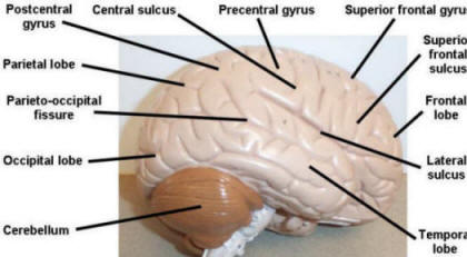
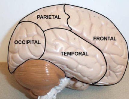
SKETCH
2
**Using the images
from this lab book and your text book, sketch an image of the
brain and identify and label the following:
Occipital lobe, Temporal lobe, Frontal lobe,
Parietal lobe
Surface Anatomy
SKETCH
3
**Using the images
from this lab book and your text book,
sketch a surface anatomy image of the brain and identify and
label the following:
Cerebellum, Cerebrum, Sulci, Gyri, Fissure
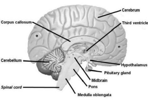
Sagittal Brain
SKETCH
4
**Using the images
from this lab book and your text book, sketch a sagittal image of
the brain and identify and label the following:
Medulla oblongata, Corpus callosum, Cerebellum,
Cerebrum, Spinal cord, Pons, Pituitary gland, Hypothalamus,
Third ventricle
Coronal Brain
SKETCH
5
**Using the images
from this lab book and your text book, sketch a coronal image of
the brain and identify and label the following:
Cerebrum, Cerebellum, Medulla oblongata, Corpus
callosum, Ventricle, Longitudinal cerebral fissure, Sulci, Gyri
FIGURE 3.3 Lateral Brain
FIGURE 3.4 Sagittal Brain
FIGURE 3.5 Superior Brain
FIGURE 3.6 Sagittal Brain
FIGURE 3.7 Sagittal Brain
FIGURE 3.8 Coronal Section of
Brain
FIGURE 3.9 Frontal Brain
FIGURE 3.10 Lateral Brain
Lobes
Human Brain
Dissection
Use the link below to
access a YOUTUBE video on the dissection of a human
brain
There are twelve pairs of nerves that emerge
from different parts of the brain and pass through openings of
the skull to parts of the head and trunk. Each pair has a name
as well as a number. Most of these nerves contain both motor and
sensory fibers and a few contain only sensory fibers. The
sensory fibers have their cell bodies in ganglia outside of the
brain. The cell bodies of the motor neurons are housed within
nuclei of the brain.
The cranial nerves are bundles of nerve
fibers that project from the brain and the brainstem. There are
twelve pairs, each known by both name and number (usually
expressed as a Roman numeral.) The cranial nerves are numbered
in the sequence of their origin from the brain, from anterior to
posterior. Sensory nerves contain only sensory (afferent)
fibers. Motor nerves contain primarily motor (efferent fibers).
Mixed nerves have significant numbers of both sensory and motor
fibers. Locate each of the twelve pairs of cranial nerves listed
here in the images provided.
HINT:
The first letter of each word in
the following sentence, or one like it, helps in memorizing
the names and numbers of the cranial nerves.
On
Old
Olympus�
Tiny
Tops,
A
Friendly
Viking
Grew
Vines
And
Hops
HINT:
The function type of each cranial
nerve can be remembered by using this
sentence:
Some
Say
�Marry
Money�,
But
My
Brothers
Say
�Bad
Business,
Marry
Money�
In this sentence,
S
indicates sensory,
M
indicates motor, and
B
indicates both
sensory and motor (mixed).
Cranial Nerves
SKETCH
6
**Using
images from the lab book and your textbook, sketch and label
a ventral image of the brain showing the cranial nerves
listed below:
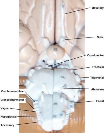
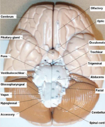
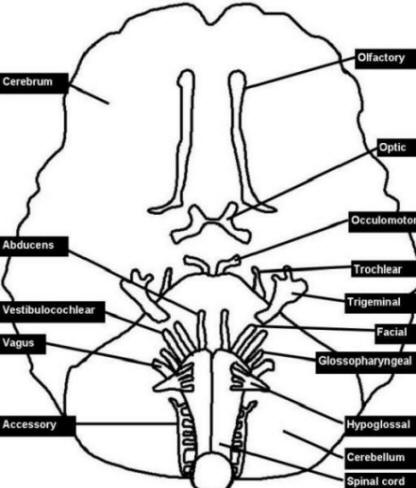
Olfactory, Optic, Occulomotor, Trochlear,
Trigeminal, Abducens, Facial, Vestibulocochlear,
Glossopharyngeal, Accessory, Hypoglossal, Vagus
This cranial nerve contains sensory fibers
for the sense of smell.
II)
Optic Nerve
This sensory nerve functions in vision. It
contains axon fibers from ganglion cells of the retina of the
eye. Some of the fibers from each optic nerve cross over to the
other side of the brain as they pass through the
optic
chiasma
.
III) Occulomotor
Nerve
This nerve emerges from the midbrain and
supplies nerve fibers to the eyelid muscles and the extrinsic
ocular muscles. The occulomotor nerve also supplies fibers of
the iris and the ciliary body. These fibers constrict the iris
and change the lens shape in
accommodation.
IV)
Trochlear Nerve
This nerve provides muscle sense and motor
stimulation of one of the muscles of the eye. This nerve emerges
from the midbrain.
V)
Trigeminal Nerve
The trigeminal nerve is the largest cranial
nerve. The trigeminal nerve is a mixed nerve but its sensory
functions are much more extensive than its motor functions.
Innervation of parts of the mouth and face are its major
functions.
VI) Abducens Nerve
This small nerve provides innervation of one
of the muscles of the eye. It is a mixed nerve in that it
provides muscle sense as well as muscular contraction.
VII) Facial Nerve
This nerve
consists of motor sensory functions. It innervates muscles of
the face, salivary glands, and taste buds of the anterior
two-thirds of the tongue.
VIII)
Vestibulocochlear
(Auditory) Nerve
This nerve goes to the inner ear. It
innervates the semicircular canals and functions in maintaining
equilibrium, while the cochlear portion is auditory in function.
IX)
Glossopharyngeal Nerve
This nerve
functions in reflexes of the heart, taste, and swallowing. Taste
buds on the back of the tongue are innervated by some of its
fibers. Efferent fibers innervate muscles controlling swallowing
and secretion from the salivary glands.
X)
Vagus Nerve
The vagus nerve supplies parts of the head
and neck with nerves and also has branches that extend down into
the chest and abdomen. It is a mixed nerve. Sensory fibers go to
the heart, external acoustic meatus, pharynx, larynx, and
thoracic and
abdominal viscera. Motor fibers pass to the pharynx, base of the
tongue, larynx, and to the autonomic ganglia of thoracic and
abdominal viscera.
XI)
Accessory Nerve
The accessory nerve innervates the
sternocleidomastoid and trapezius muscles. The cranial portion
of the nerve innervates the pharynx, upper larynx, uvula, and
palate.
XII) Hypoglossal
Nerve
This nerve innervates several muscles of the
tongue. It contains both afferent and efferent fibers.
Click the play button to
view the movie to the right on identifying the cranial nerves.
An alternative view
for this animation is given below:
Evidence of nerve damage could mean a
peripheral lesion in the nerve or a central lesion in the
brainstem. An instruction sheet and data table to perform the cranial nerve
function tests can be accessed from the SKETCHES TABLES
CHARTS Form. Conduct
the tests on two willing subjects or lab partners and indicate
in the subject column with a �+� or �-� sign the
results of the test. A �+� sign would indicate that the
test subject has passed the test and is able to perform or
produce the results asked for.
TABLE
**Print off the cranial nerve function table and
record your results on the sheet.
The Cranial Nerve
Function Table is available by printing out the Sketches
Tables Charts Form
Click the play button to
view the movie to the right on techniques for testing for normal cranial nerve
function.
An alternative view
for this animation is given below:
**At
the following sites, perform the memory tests as indicated.
Access a table that will show the results of your tests for
each by using the link given below.
The Short Term Memory Table is available by
printing out the Sketches Tables Charts Form
The cerebral hemispheres of the brain are
divided into a right hemisphere and a left hemisphere. Each
hemisphere appears to be specialized for certain behaviors. The
left brain may be more dominant for calculations, math and
logical abilities while it appears that the right brain is
dominant for spatial abilities, face recognition, visual imagery
and music. The right side of the brain is
intuitive, while the left side of the brain is logical.
Of course, these are generalizations and in normal people, the
two hemispheres work together. They are connected, and share
information through a thick band of nerve fibers called the
corpus callosum. Much of what we know about the right and left
hemispheres comes from studies in people who have had the corpus
callosum split. This surgical operation isolates most of the
right hemisphere from the left hemisphere. This type of surgery
is performed in patients suffering from epilepsy. The corpus
callosum is cut to prevent the spread of the "epileptic seizure"
from one hemisphere to the other.
Even though both hemispheres of
the brain have independent functions, an individual benefits
from the integration of the processing of information performed
by each side. The hemisphere best suited to perform the
processing will process information; this allows an individual
greater understanding and learning potential of the situation
that initiated the brain processing information.
The left side of the brain controls muscles on
the right side of the body and the right side of the brain
controls muscles on the left side of the body. In general,
sensory information from the right side of the body crosses over
to the left side of the brain and information from the left side
of the body crosses over to the right side of the brain. Thus,
damage to one side of the brain will affect the opposite side of
the body.
The left side of the brain is dominant for
language in 95% of right handers. Even in 60-70% of
left-handers, the left side of brain is used for language.
Neurologists observe that people who have had damage to a
particular area on the left
side of the brain had speech and language problems. In
most cases people with damage to these areas on the right side
did not have any language problems. The two language areas of
the brain that are important for language are Broca's area and
Wernicke's area.
Research has shown most people
have a dominant side of the brain. Individuals who are
predominately left sided tend to be more verbal, analytical, and
problem solvers; while individuals who are predominately right
sided tend to be artsy, good with math, and are more visual in
nature. Dominance goes into affect when thinking becomes
increasingly more complex. Although each hemisphere has its own
set of functions in information processing and thinking,
research data supports the notion these functions are not
exclusive to one hemisphere.
Which side of your brain is dominant? Use the
questionnaire provided below to access a test to find your
BRAIN
DOMINANCE. The Brain Type Test will
determine which half is your dominant half, and to what degree.
The test consists of 20 questions. After completing the test, you will be
given instructions on how to calculate your
left and right brain score. Enter your BRAIN DOMINANCE SCORE and list
five general dominant
traits for that side of the brain in the BRAIN HEMISPHERE DOMINANCE DATA TABLE.
The web site listed below presents animations
and discussion on the physiology of neurons in the brain. The
animations specifically illustrate the action of
neurotransmitters in the brain. Become familiar with the normal
functioning that is illustrated, as well as reading the
information provided for each of the slides. As you view the
animations, you may want to take a few notes to help you answer
questions about the exercise. The sites will give a menu of
options for you to view. The section we are interested in
studying falls under the "IN THE BRAIN" listing. There
are four parts to the study:
1) How Brain Cells Communicate
2) How Cocaine Works in the Brain
3) How Alcohol Works in the Brain
4) How Opiates Work in the Brain
QUESTIONS **View
the "How Brain Cells Communicate" animation. Next you will
view the other parts of the study that are listed above by
utilizing the links given on the site. When you are
finished, you will need to answer questions 1-8 that are
given below.
1)
What role does GABA play in the nervous
system? Please be descriptive. 2)
Describe the effect that alcohol has
on the normal functioning of GABA receptors. 3)
Think of a time when you witnessed
someone under the influence of alcohol. What physical behaviors
are characteristic of intoxication? How might you explain these
characteristics using the images you just viewed?
4) In light of these facts, why is it a bad idea
to drive while intoxicated? How might
alcohol affect one's driving ability?
5) Describe how cocaine�s ability to block
reuptake pumps for dopamine causes an
intense feeling of euphoria.
6) Why does one need to take higher and higher
doses of cocaine in order to feel the same sense of pleasure
from the drug?
7) Describe normal dopamine function.
8) How are the natural levels of dopamine
altered by use of opiates?
The Brain Physiology
Questions are available by printing out the Questions
Form
Jokes about men vs women are probably as old as language
itself, but is there any science behind the notion that men and
women have fundamentally different brains? It is thought that
everybody�s brain has a gender. It�s nothing to do with sexual
orientation or your actual gender, but you�ve almost certainly
got either a systemising (male) brain, or an empathising
(female) brain. They�re wired to think, feel and react
differently, but it�s perfectly common for men to have
empathising (female) brains and women to have systemising (male)
brains.The type of brain you have will influence your feelings,
behaviour, talents and weaknesses. We enlisted a set of
volunteers, some of whom do jobs stereotypically associated with
their gender and some who do the opposite.Dr Anne Moir believes
our life paths might not be down to choices and social
conditioning, but in fact may be beyond all control. They could
be a result of the gender of our brains. In a surprising twist,
simply measuring your ring finger can reveal much about the
inner workings of your mind. It is thought that the longer your
ring finger in relation to your index finger, the more
testosterone you were exposed to in the womb. Dr Christian�s
brain is somewhere in the middle, with his index and ring
fingers being more or less equal in length.Good hand-eye
coordination is considered to be a systemising (male) trait, as
is being able to visualise shapes in 3D. Conversely those with
empathising (female) brains are much better at deciphering
expressions in other people. They are better at judging emotions,
joining groups and short term memory.
Perform the BRAIN GENDER ID
TEST to find if you have a male or
female oriented brain. You will find the Questionnaire in the
Sketches Tables Charts Form
TABLE **Fill
out the Questionnaire provided in the Sketches Tables Charts
Form and submit with your
lab report
The Brain Gender
Questionnaire is available by printing out the
Sketches Tables Charts Form
Biology of Love (optional) Interesting discussions of the power of hormones and
neurotransmitters in the brain. There are no
requirements or assignments to view these two videos. You
may view them at your own option.
Click the play button to
view the movie to the right discussing the actions of
brain in love. (Part 1)
An alternative view
for this animation is given below:
The presence of electrical
current in the brain was discovered by an English physician,
Richard Caton, in 1875. It was not until 1924 that Hans
Berger, a German neurologist, used his ordinary radio
equipment to amplify the brain's electrical activity so that
he could record it on graph paper. Berger noticed that
rhythmic changes (brain waves) varied with the individual's
state of consciousness. The various regions of the brain do
not emit the same brain wave frequency simultaneously. An EEG electrode placed on
the scalp would pick up many waves with different
characteristics. This has presented a great deal of
difficulty to researchers trying to interpret the large
amount of data they receive from even one EEG recording.
Brain waves have been categorized into four basic groups:
Alpha,
Beta, Theta,
and Delta
waves. Although none of these waves is ever emitted alone,
the state of consciousness of the individual may make one
frequency more pronounced than the others.
You may have seen
doctors in hospitals or on television taking EEG
readings of the natural and ongoing electrical activity
of a person's brain. This activity is produced by all of
us all the time and it varies according to what kind of
activity we are engaged in. It can be recorded by
sensors that are gently placed on the head. A geodesic
sensor net, which looks a bit like a hairnet with lots
of little sponges attached to it can be used to acquire
this electrical activity. The net contains 64 sensors,
which are all sewn together.
Brain waves are obtained
from a special analysis of EEG. These brain waves show us
the brain's response to a particular stimulus or external
event, such as a picture or sound. Brain activity before,
during, and after a stimulus presentation is recorded. This
allows us to observe where, when, and how the brain responds
to a given stimulus. Any physiological
investigation of the brain can emphasize and expose only
a very minute portion of its activity. Higher brain
functions, such as consciousness and logical reasoning,
are extremely difficult to investigate.
It is obviously much
easier to do experiments on the brain�s input-output
functions, some of which can be detected with
appropriate recording equipment. Still, the ability to
record brain activity does not necessarily guarantee an
understanding of the brain. Certain characteristics of
brain waves are known. They have a frequency of 1 to 30
hertz (Hz) or cycles per second, a dominant rhythm of 10
Hz and an average amplitude (voltage) of 20 to 100
microvolts (uV).
Frequency
is the number of times a wave repeats itself
within a second. It can be compared to the
frequencies that you tune into on your radio. If
any of these frequencies are deficient,
excessive, or difficult to access, our mental
performance can suffer.
Amplitude
represents the power of electrical impulses
generated by brain. A wave can be of high or low
amplitude (voltage) and high or low frequency
(regularity).
The first of the brain
waves to be described by scientists were the
Alpha Waves (or
alpha rhythm). Alpha waves have an average frequency
range of 8 to 13 Hz and are produced when the individual
is in a relaxed state with the eyes closed. Alpha block,
suppression of the alpha rhythm, occurs if the eyes are
opened or if the individual begins to concentrate on
some mental problem or visual stimulus. Under these
conditions, the waves decrease in amplitude but increase
in frequency. Under conditions of fright or excitement,
the frequency increases still more. Beta Waves,
closely related to alpha waves, are faster, 14 to 30 Hz
and have a lower amplitude. They are typical of the
attentive or alert state. Very large (high-amplitude)
waves with a frequency of less than 4 Hz that are seen
in deep sleep are Delta Waves.
Theta Waves are
large, abnormally contoured waves with a frequency of 4
to 7 Hz. Although theta waves are normal in children,
they represent emotional problems or some sort of neural
imbalance in adults. Gamma Waves,
are brain waves larger than 30 Hz. These waves
predominate during periods of times we are �thinking�.
In normal adults who
are awake, the EEG shows mostly Alpha waves and Beta
waves. In abnormal adults the EEG shows sudden bursts of
electrical activity (spikes) or sudden slowing of brain
waves. These abnormal discharges may be caused by a
brain tumor, infection, injury, stroke, or epilepsy.
When a person has epilepsy, the location and exact
pattern of the abnormal brain waves may help determine
what type of epilepsy or seizures the person has. Keep
in mind that in many people with epilepsy, the EEG may
appear completely normal between seizures. A disorder affecting
the entire brain, such as drug intoxication, certain
infections, or metabolic disorders that upset the
chemical balance in the body, including the brain, may
produce abnormal brain waves. In these abnormalities the
EEG shows delta waves or an excess of theta waves in
adults who are awake. These results may indicate brain
injury.
If the EEG shows no
electrical activity in the brain (a "flat" or
"straight-line"). This indicates that brain function has
stopped, which is usually caused by lack of oxygen or
blood flow inside the brain. In some cases, severe
drug-induced sedation can produce a flat EEG. This state
also can be seen in status epilepticus after a
significant amount of medication is given to control the
seizure. A person who has a flat EEG for more than 6
hours is usually considered brain dead, unless heavily
sedated with medications. Brain waves change with
age, sensory stimuli, brain pathology or disease.
Glucose deprivation, oxygen poisoning and sedatives all
interfere with the rhythmic activity of brain output by
disturbing the metabolism of the neurons. Sleeping
individuals and patients in a coma have EEGs that are
slower (or lower frequency) than the alpha rhythm of
normal adults. Fright, epileptic seizures, and various
types of drug intoxication are associated with faster
brain activity. Impairment of brain function is
indicated by neuronal activity that is either too fast
or too slow.
We are now ready to
begin the brain wave portion of the lab. You will be
assigned a subject who will perform various mental
activities for you. You will be using a device which
will be able to record the brain waves of your
subject while they are performing the mental tasks.
You will need to record the mental activities
attempted and sketch and identify the brain waves
which are recorded. You will also need to answer
questions on your data collected.
I
n order
to perform the lab you will need to Link to the link
given below. For this portion of the lab you will need to
provide a Data Table for Brain Waves and answers to
questions on Brain Waves.
1) Describe the difference between
amplitude and frequency in regards
to brain waves. 2) Describe the brain waves of an
individual who is �brain dead�. 3) List the dominant brain wave we
would find in individuals performing
the following activities:
a)
Sleeping b) Under Stress c) Relaxing with eyes open d) Concentrating
The Brain Wave Questions are
available by printing out the
Sketches Tables Charts Form
**Utilizing
the site listed below, perform the brain surgery as directed
by the simulation. After finishing the simulation answer the
following questions.
Questions on Virtual Brain Surgery
1)
Why does the hair need to be shaved from the head?
2)
Why are so many scrubbings and drapes necessary?
3)
Why is a saline solution used during drilling through
the skull?
4)
What portion of the brain is being stimulated?
5)
What condition is the patient exhibiting?
The Virtual Brain Surgery Questions are
available by printing out the Sketches Tables Charts
Form
View the following three
tutorials of Diseases and Condtions of the Brain and produce
a chart for each which summarizes the main points for the
tutorial. You need to use the charts that are provided in
the links below.