|
|
AP 2 Lab Manual |
If you disable the "Active Content" in your browser you may not be able to view the animations or videos supplied in this lab. If prompted you should "Allow Blocked Content". |
LAB 1
NERVES

|
CONTENTS
|
What Do I Have To Hand In For This Lab? Sketches
Chart Questions
Cover Page |
|
FORMS REQUIRED FOR THIS LAB |
|
| MS WORD | |
| Lab 1 Cover Page (not available) | Lab 1 Cover Page |
| Sketches | Sketches |
| Tables and Charts | Tables and Charts |
| Questions | Questions |
| Access each of the listed documents above and print them off. When you submit your lab report you will need to compile all of the documents listed above, stapled together in the order listed in the table above. Sketches must be performed free hand (not traced or copy and pasted). Sketches must be performed using the printed links as given above. You are not allowed to perform the sketches on blank sheets of paper or lined sheets of paper. Sketches performed without using these forms above will not be accepted. You can use the MS WORD links to access the questions, tables and charts in order to input your values or answers electronically and then print them off when finished to include with your lab report. Alternatively you can print the questions, tables and charts forms out and input your values or answers by hand. The PDF file format will not allow you to input values or answers electronically. Please collate and order the pages in your lab report in the order they are listed in the table above. The cover page is only available using the PDF file format. |
Nerve Tissue
Nerve cells can be divided into two main categories: 1) Neurons, 2) Neuroglia. Each of these type of cells have different structures and different functions. Neurons are the types of nerve cells which are able to carry nerve impulses. Neuroglia also called glial cells, have an important function in various supporting roles of the neurons. Neurons and neuroglia are found not only in the brain and spinal cord but in the peripheral regions of the body as well.
Neurons are the structural units of nervous tissue. They are highly specialized to transmit nerve impulses from one part of the body to another. Although neurons differ structurally, they have many identifiable features in common.
|
All have a cell body from which slender processes extend.
Although neuron cell bodies are typically found in the brain and
spinal cord in clusters called nuclei, occasionally they reside
in ganglia (collections of neuron cell bodies outside the CNS).
They make up the gray matter of the nervous system. Neuron
processes running through the CNS form tracts of white matter;
outside the CNS they form the peripheral nerves.
|
|

| According to the traditional scheme, neuron processes that conduct electrical currents toward the cell body are called dendrites, and those that carry impulses away from the nerve cell body are called axons. Neurons have only one axon but may have many dendrites, depending on the neuron type. |
|

In general, a neuron is excited by other neurons when their axons release neurotransmitters close to its dendrites or cell body. The electrical current generated travels across the cell body and down the axon. The axon begins at the cell body and ends in many small structures called synaptic knobs. These knobs store the neurotransmitter chemical in tiny vesicles. Each synaptic knob is separated from the cell body or dendrites of the next neuron by a tiny gap called the synaptic cleft. Thus, although they are close, there is no actual physical contact between neurons. When an impulse reaches the synaptic knobs, some of the synaptic vesicles rupture and release neurotransmitter into the synaptic cleft. The neurotransmitter then diffuses across the synaptic cleft to bind to membrane receptors on the next neuron, initiating the nerve impulse.
|
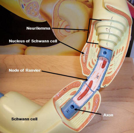
Many drugs can influence the transmission of impulses at synapses. Some, like caffeine, are stimulants which decrease the receptor neuron�s threshold and make it more irritable. Others block transmission by binding competitively with the receptor sites or by interfering with the release of neurotransmitter by the synaptic knob. As might be anticipated, some of these drugs are used as painkillers or tranquilizers. Most long nerve fibers are covered with a fatty material called myelin, and such fibers are referred to as myelinated fibers. Axons in the peripheral nervous system are typically heavily myelinated by special cells called Schwann cells, which wrap themselves tightly around the axon. This wrapping is the myelin sheath. The peripheral part of the Schwann cell and its plasma membrane is referred to as the neurilemma. The myelin sheath is formed by many individual Schwann cells; thus gaps in the sheath are formed called nodes of Ranvier. Within the brain and spinal cord, myelination is accomplished by glial cells called oligodendrocytes. These glial cell sheaths do not exhibit the neurlolemma seen in fibers myelinated by Schwann cells. Myelin insulates the fibers and greatly increases the speed of neurotransmission by neuron fibers.
|
|
|
|||
|
|
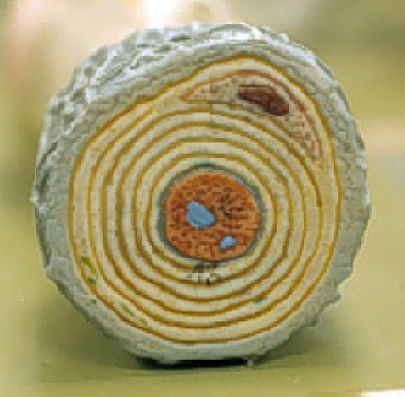
FIGURE 1.5 Cross section of Axon
|
||

|
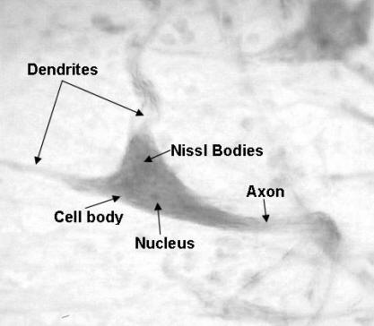
2) Spinal Cord Smear The image to the right is a prepared slide of teased nerve tissue, which has large, easily identifiable neurons. The neuron represented here was photographed under high power(400X). Sketch the neuron cell and label the following: Cell Body, Nucleus, Nissl Bodies, Dendrites, Axon
|
|
3) Neuron
Anatomy
Quiz
|
|
|
The Neuron Anatomy Quiz is available by printing out the Sketches Tables Charts Form |
|

Use the images below and images in your text book in performing the sketches listed below
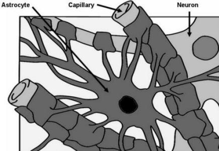
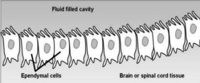
Sketch the following neuroglia types and indicate their functions within the CNS:SKETCH 3
**
Ependymal Cells, Oligodendrocytes, Astrocytes, Microglia
|
|
|

|
|
|

|
Click the play button to
view the movie to the right on the importance of glial
cells. An alternative view for this animation is given below: http://www.youtube.com/watch?v=itinDmvDEqw |
|
Neurons may be classified on the basis of structure or of function. Basis of Structure Structurally, neurons may be defined according to the number of processes attached to the cell body. In unipolar neurons, one short process extends from the cell body. Functionally, only the most distal portions of the peripheral process act as dendrites; the rest acts as an axon along with the central process. Most neurons that conduct impulses toward the central nervous system are unipolar. Bipolar neurons have two processes (one axon and one dendrite) attached to the cell body. This neuron type is quite rare, typically found only as part of the eye, ear, and olfactory mucosa. Many processes issue from the cell body of multipolar neurons, all classified as dendrites except for a single axon. Most neurons in the brain and spinal cord and those, whose axons carry impulses away form the CNS fall into this multipolar category. Basis of Function
Neurons carrying impulses
from the sensory receptors in the internal
organs or in the skin are termed sensory, or
afferent, neurons. The dendritic endings of
sensory neurons are equipped with specialized
receptors that are stimulated by specific
changes in their immediate environment. The cell
bodies of sensory neurons are found in ganglion
outside the CNS, and these neurons are typically
unipolar. Neurons carrying activating impulses
from the CNS to the viscera or body muscles and
glands are termed motor, or efferent, neurons.
Motor neurons are for the most part multipolar.
Their cell bodies are always located within the
CNS and they are multipolar neurons
structurally. |
|
|
|


|
|
6) Structure of a Nerve
A nerve is a bundle of neuron fibers wrapped in connective tissue coverings and that extends to and from the CNS and visceral organs or structures such as skeletal muscles, glands, and skin. Within a nerve, each fiber is surrounded by a connective tissue sheath called an endoneurium, which insulates it from the other neurons adjacent to it. Groups of fibers are bound by a coarser connective tissue, called the perineurium, which form bundles of fibers called fascicles. Finally, all the fascicles are bound together by a tough, fibrous connective tissue sheath called the epineurium. The entire bundle forms the cord-like nerve. Blood vessels and lymphatic vessels serving the fibers also travel within a nerve. Nerves are classified according to the direction in which they transmit impulses. Nerves carrying both sensory (afferent) and motor (efferent) fibers are called mixed nerves. All spinal nerves are mixed nerves. Nerves that carry only sensory processes are referred to as sensory, or afferent, nerves. Some of the cranial nerves are pure sensory nerves, but the majority are mixed nerves. The ventral roots of the spinal cord, which carry motor fibers, are considered motor or efferent nerves. A prepared cross section of a peripheral nerve is shown below. Use these figures to sketch the image asked for below.
|
|

|
|
|
|
|


**Answer the following questions (at the bottom of the page) about nerve impulses.
This site has an animation of a nerve impulse through a nerve cell.
|
|
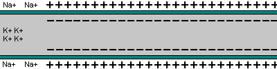
The diagram seen here represents an axon terminal on the left and a dendritic process on the right separated by a synaptic cleft. When an impulse reaches the end of an axon, it triggers the formation of synaptic vesicles at that terminal. Synaptic vesicles are specialized vacuoles that contain neurotransmitters such as acetylcholine. The vesicles transport the neurotransmitters to the end of the axon and release them into the synaptic cleft. These neurotransmitters attach to receptor sites on the cell membrane of the receiving neuron. When enough receptor sites are filled, the firing threshold of the receiving neuron is reached and a depolarization event is triggered.
Before the neuron depolarizes, it is held steady in its resting potential. This potential, which is achieved by maintaining a relatively high concentration of sodium ions outside of the cell membrane, represents an approximately -70 milli-volts discrepancy between the negatively charged interior and positively charged exterior. As neurotransmitters attach to the receptor sites and overcome the firing threshold, small molecular gates open along the cell membrane allowing the sodium ions to rapidly flood the neuron. This sudden change in polarity from the influx of positive ions triggers an action potential that moves like a wave down the axon triggering another nerve, muscle cell, etc.
At this point, a different series of molecular gates open which allows potassium ions to rush out of the neuron. The potassium ions, which have a positive charge as well, create a negatively charged cell interior by their absence. This event stops the depolarization process. The sodium ions are pumped more slowly to the cell exterior by active transport, resulting in the fully restored resting potential once again.
|
Click the play button to
view the movie to the right describing the action
potential in a nerve axon. An alternative view for this animation is given below: |
QUESTIONS
**Neuron Function Questions
1) Describe some differences between the axon and dendrite of a cell.
2) Where are the synaptic vesicles formed?
3) What is the name of the space between two interacting neurons?
4) What is the collective name of the chemicals stored in the vesicles?
5) Where is the highest concentration of Na+ (sodium) ions, inside the axon or outside?
6) How many milli-volts is a normal resting potential
The Questions are available by printing out the Questions Form
8) Nerve Disorders
View the following three tutorials of Diseases and Conditions of the Nervous System and produce a chart for each which summarizes the main points for tutorial. You need to use the Charts that are supplied in the links below.
CHARTS
The Nerve Disorders Chart is available by printing out the Sketches Tables Charts Form
END LAB 1