|
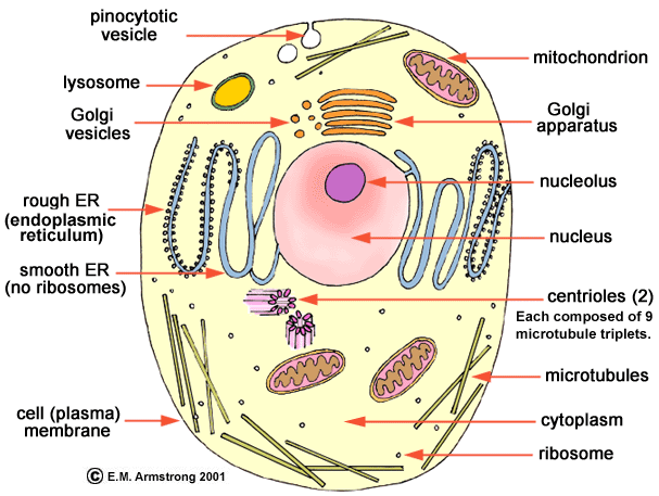
Animal Cell Illustration With Hyperlinked Labels
Golgi Vesicle: A membrane-bound body that forms by "budding" from the Golgi apparatus. It contains proteins (glycoproteins), such as digestive enzymes, and migrates to the cell (plasma) membrane. Golgi vesicles fuse with the cell membrane and discharge their contents into the exterior of the cell through a process called exocytosis. Some Golgi vesicles become lysosomes which are involved in intracellular digestion. Pinocytotic Vesicle: A membrane-bound vacuole formed by a specific type of endocytosis called pinocytosis. The plasma membrane invaginates (pinches inwardly) to form a vesicle that detaches and moves into the cytoplasm. Macromolecular droplets and particles up to 2 micrometers in diameter enter the cell within these pinocytotic vesicles. Larger particles (including bacteria) enter special white blood cells (phagocytes) through a form of endocytosis called phagocytosis. The Amoeba is a unicellular protist that ingests food (including algal cells) by phagocytosis. Lysosome: A membrane-bound organelle containing hydrolytic (digestive) enzymes. Lysosomes originate as membrane-bound vesicles (called Golgi vesicles) that bud from the Golgi apparatus. They are primarily involved with intracellular digestion. Lysosomes fuse with vesicles (small vacuoles) formed by endocytosis. The contents of these vesicles are digested by lysosomal enzymes. Autodigestion by lysosomes also occurs during embryonic development. The fingers of a human embryo are webbed initially, but are separated from each other by lysosomal enzymes. Cells in the tail of a tadpole are digested by lysosomal enzymes during the gradual transition into a frog. Mitochondrion: Membrane-bound organelle and the site of aerobic respiration and ATP production. Energy from the step-by-step oxidation of glucose (called the Krebs or citric acid cycle) is used to produce molecules of adenosine triphosphate (ATP). The Krebs cycle starts when a 2-carbon acetyl group combines with a 4-carbon group to form a 6-carbon citrate. Including glycolysis (which occurs outside the mitochondria), a total of 38 ATP molecules are generated from one molecule of glucose. Endoplasmic Reticulum: A complex system of membrane-bound channels extending throughout the cytoplasm of cells. Like the emergency room of a hospital, the endoplasmic reticulum is often abbreviated as ER.
Ribosome: Organelle site of protein synthesis. The ribosome is composed of large and small subunits separated by a central groove. A strand of messenger RNA (m-RNA) fits into the groove and the ribosome moves along the m-RNA in a 5' to 3' direction. Molecules of cloverleaf-shaped transfer-RNA (t-RNA), each with a unique amino acid, temporarily attach to the m-RNA at the ribosome in a process called translation. Transfer-RNA anticodons hook up with m-RNA codons and the amino acids bond together by dehydration synthesis. As the ribosome moves toward the 3' end of the m-RNA strand, the amino acid chain (polypeptide) grows longer and longer. Finally the completed polypeptide leaves the ribosome site and moves away to become a protein utilized within the cell or secreted from the cell. The simplified animated gif images below illustrate this remarkable process. A series of several ribosomes moving along the same m-RNA strand is called a polyribosome. Ribosomes are composed of ribosomal RNA and they are not membrane-bound. They occur in prokaryotic as well as eukaryotic cells. In eukaryotic cells, ribosomal RNA is synthesized in the nucleolus. The large and small subunits of ribosomes are synthesized by specific genes. One gene in the nucleolus codes for the smaller subunit of the ribosome. The gene is called SSU rDNA or small subunit ribosomal DNA. Base sequences from this gene are sometimes used to compare taxa at the species level. The results from comparative DNA studies using mitochondrial and chloroplast DNA are illustrated in computer generated evoltionary trees called cladograms. Nucleolus: Dark-staining body within the nucleus where ribosomal RNA is synthesized. Plant nuclei in onion root tip cells may have several nucleoli. Nucleus: Membrane-bound organelle containing chromatin, a term applied to all the chromosomes collectively when they are in a tenuous (threadlike) stage. During the prophase of mitosis, the chromosomes become shorter and thicker, and appear as distinct doubled bodies called "chromosome doublets." Cell (Plasma) Membrane: The living membrane that surrounds the cytoplasm of all cells. It is composed of a phospholipid bilayer with embedded glycoproteins. In the "sandwich model" the two phospholipid layers are sandwiched between two layers of protein. The membranes of organelles are also composed of a phospholipid bilayer, including vacuoles, nuclei, mitochondria and chloroplasts. [Riubosomes are not membrane-bound.] Embedded glycoproteins in plasma membranes include membrane transport "carrier molecules" and cell recognition antigens. The plasma membrane is permeable to water molecules by osmosis, but not to other molecules and ions by simple diffusion. Ions pass through the plasma membrane via carrier molecules by active transport and facilitated diffusion. Active transport requires ATP. Centrioles: Nonmembrane-bound organelles that occur in pairs just outside the nucleus of animal cells. Each centriole is composed of a cylinder or ring of 9 sets of microtubule triplets with none in the middle (9 + 0 pattern). During cell division a pair of centrioles moves to each end of the cell, forming the poles of the mitotic spindle. Centrioles also give rise to basal bodies that control the origin of cilia and flagella in motile cells of protists. In cross section, flagella and cilia have 9 sets of microtubule doublets surrounding a pair of single microtubules in the center (9 + 2 pattern). This characteristic pattern also occurs in motile cells of higher organisms, such as human sperm. Microtubules: Protein filaments composed of a polymer called tubulin. The centrosome of animal cells (including a pair of centrioles and radiating aster) are composed of microtubules. Microtubules are involved in cell movement, cell shape and the formation of mitotic spindles during cell division (mitosis). Some cancer chemotherapy drugs cause the dissolution (depolymerization) of tubulin in microtubules, thus destroying mitotic spindles and effectively stopping cell division in tumor cells. Cytoplasm: All the contents of a cell within the plasma membrane. The nucleus and its contents (nucleoplasm) are generally excluded from the cytoplasm. The semifluid medium between the nucleus and the plasma membrane is called cytosol.
|
